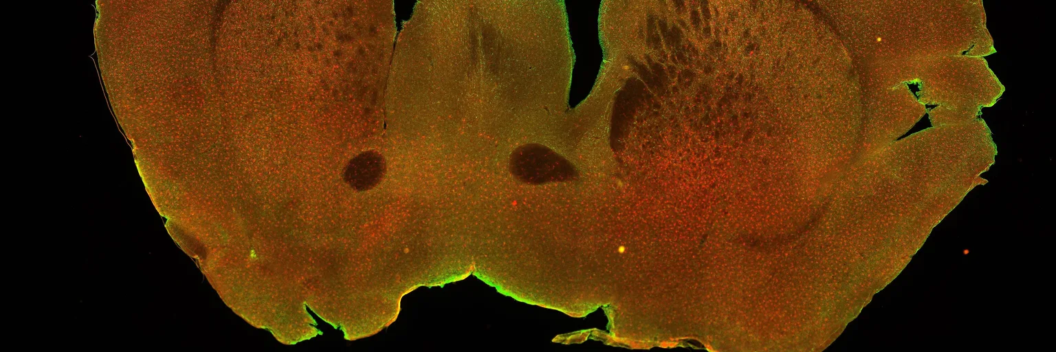
Biology studied cells in snapshots; live 3D film shows what we missed
Filming the inside of a living cell in 3D turns biology from a photo album into cinema, as long as we can prove the camera doesn't change the plot.
Photo: ZEISS Microscopy from Germany / Wikimedia Commons (CC BY-SA 2.0)
Imagine trying to understand a city by looking at a handful of aerial photos, each taken on a different day, some of them after the city had been frozen and sliced open. That, more or less, is how a great deal of cell biology has worked. A wave of new microscopes now lets researchers watch the inside of a living cell in three dimensions, and we think the right reaction is not "how pretty" but "how much did we assume?"
Several labs are racing to film living cells
Gizmodo reports on a new microscopy technique that records the interior of a cell in three dimensions fast enough to turn a sequence of images into a video. Elsewhere, University of Tokyo researchers have developed a high-speed method that makes "videos" of the dynamic processes inside cells, according to Phys.org. Stanford has a microscope that lets scientists watch structures inside living cells move and interact in real time. And a recent news-medical.net piece covers a label-free technique based on mid-infrared photothermal microscopy that films cell activity in 3D.
Most of what we reviewed is press-level summaries, so we can't vouch for the peer-review status of every result. One we can place: the Stanford approach, called Interferometric Image Scanning Microscopy, was published on February 27, 2026 in Light: Science & Applications, with a lateral resolution of about 120 nanometers.
Motion is data, not decoration
A still image of a cell answers "what is where?" A film answers "what happens next, how fast, and who touches whom?" Those are different scientific questions, and the second is closer to what a cell actually does. A structure that looks like an isolated blob in a frozen frame may turn out to be a transient visitor. Two organelles that appear to sit apart may in fact meet constantly. In our view, that is where the real payoff lies: not sharper pictures, but hypotheses that can only be formed once timing is visible.
Will textbooks need redrawing? Partly, and we'd bet on the diagrams first. Those tidy cutaways with every compartment neatly labeled and static are a convention born of the tools available, not a claim about how cells behave. We expect the facts in those books to survive far better than the illustrations. Anyone promising that "everything we knew was wrong" is selling a headline. What changes is emphasis: dynamics move from footnote to main text.
The camera is not innocent
Here is the part we would not let anyone skip. Every way of looking at a living thing risks changing it. Fluorescent labels add molecules to the cell, and intense illumination can stress it. That is why the label-free pitch matters. Stanford's team says its microscope blends two imaging approaches and allows nearly unlimited observation time without labels, and another Stanford page notes a resolution of about 120 nanometers without fluorescent labels. If that holds up, it removes one big source of doubt.
The other source is software. UC San Diego engineers say their AI-enhanced method delivers sharper real-time video inside live cells without misleading image artifacts. That claim deserves applause and skepticism in equal measure. When an algorithm cleans up an image, the question is always whether it reveals structure or invents it, and the best answer is independent validation against other methods, by groups with no stake in the result.
So our checklist for any "we filmed a living cell" announcement is short. Do the cells behave normally while being watched, and for how long? Is the result reproduced in different cell types and different labs? Does the footage match what older techniques show where they overlap? The coverage we reviewed does not answer these, and we won't pretend it does.
Our verdict
This is one of the more underrated stories of the season, precisely because it lacks a single dramatic discovery. It is a change in the kind of evidence biology can collect, and those changes tend to matter more than any one finding. Our recommendation to readers: enjoy the videos, but judge each technique by how well it shows it isn't altering the cell it films. The best microscope here will not be the prettiest one. It will be the one whose footage still looks the same when someone else runs the experiment.
No topics yet: start the first one.
More stories
Vitamin A and vision: a mechanism update, not a prescription
A Johns Hopkins study on how sharp vision is built before birth reminds us how much we still don't know about the eye, and why it shouldn't be sold as a pill.
Quantum sensors could replace animal tests for drug safety, if they prove themselves
NASA and the NIH want quantum sensors to read organ-on-a-chip models without animals. It's a smart bet, but only if the readings are checked against real physiology.
Revived mouse brain tissue is real progress, but not a mind
German researchers kept mouse brain tissue functional after vitrification. That is a genuine preservation advance, and cryonics hype should not borrow its credibility.
Cold sore virus and the brain: a clue for Alzheimer's debate, not a verdict
A mouse study suggests herpes simplex reprograms the brain's immune cells. It is a useful mechanistic hint for the Alzheimer's debate, but it doesn't show cold sores harm your brain.